Authored by Vince Murdock, moyamoya survivor and the Miles for Moyamoya team. Sources are cited inline and listed in full at the page bottom. Last updated: 25 May 2026.
The disease takes its name from how it looks on a brain scan, and the science of why it looks that way.
Moyamoya disease is a chronic, progressive narrowing of the terminal internal carotid arteries and their branches at the base of the brain. As the main arteries close, the brain compensates by recruiting a tangled network of small, fragile collateral vessels. On a cerebral angiogram, that network looks hazy and wispy, like a drifting cloud of cigarette smoke. Japanese neurosurgeons Jirō Suzuki and Akira Takaku coined the term moyamoya (もやもや, "puff of smoke") in their seminal 1969 description.
The narrowing is not caused by cholesterol or inflammation. Under the microscope, the affected arteries show thickening of the inner layer (intima) and thinning of the muscular middle layer (media). The process is slow, progressive, and currently irreversible. The collateral vessels the brain grows in response are fragile and prone to both ischemic stroke (too little blood) and hemorrhagic stroke (vessel rupture) (AHA/ASA Scientific Statement, 2023).
Doctors distinguish between two forms. Moyamoya disease is the primary, idiopathic form with no underlying cause. Moyamoya syndrome describes the same imaging appearance occurring alongside another condition such as sickle cell disease, neurofibromatosis type 1, Down syndrome, prior cranial radiation, or certain autoimmune diseases (NINDS, 2026).
The progressive narrowing of critical arteries forces the brain to build fragile backup blood vessels that cannot sustain healthy function.
In a healthy brain, the internal carotid arteries deliver oxygen-rich blood through the Circle of Willis at the brain's base. In moyamoya disease, these arteries progressively narrow because of smooth-muscle thickening in the vessel wall. The RNF213 gene on chromosome 17q25.3 is the principal susceptibility gene, especially in East Asian populations. The narrowing is irreversible and worsens over time, gradually cutting off the brain's primary blood supply.
As the main arteries close, the brain compensates by growing a network of tiny collateral blood vessels around the blockage. These thin-walled vessels are insufficient and prone to rupture, which is why moyamoya causes both ischemic and hemorrhagic strokes.
The result is a brain slowly starving of oxygen. Without surgical intervention, the cumulative recurrent-stroke risk reaches roughly 40% at 5 years, climbing higher in bilateral disease. Children may experience strokes triggered by crying, exercise, or hyperventilation. Adults are more likely than children to present with brain hemorrhage.
Moyamoya can affect anyone but follows recognisable patterns by geography, sex, and age.
Japan reports a prevalence of around 3.16 per 100,000. US incidence is about 0.086 per 100,000, roughly ten times lower. Asian Americans are affected approximately four times more often than white Americans (Epidemiology of moyamoya, PMC4747069).
US data show women account for around 72% of cases. Japanese national registry data report a female to male ratio of roughly 1.8:1 (Japan National Registry, Stroke 2019).
The disease shows two incidence peaks: ages 5 to 10 (paediatric, mostly ischemic strokes) and ages 30 to 50 (adult, with a larger share of hemorrhagic strokes). Vince Murdock was diagnosed at 30, squarely in the adult peak.
Moyamoya has a 62% misdiagnosis rate, with an average 5.28 years from first symptom to correct diagnosis. Children almost always present with ischemic events. Adults can present with either ischemic or hemorrhagic stroke, and are about 7x more likely than children to bleed.
The 62% misdiagnosis rate reflects low clinical awareness outside specialist centres. For a full breakdown of warning signs and red flags, see the full symptom guide.
62%
MISDIAGNOSED
In the largest Caucasian moyamoya series, more than three in five patients were initially given the wrong diagnosis. Mean delay to correct diagnosis: 5.28 years.
See the warning signs →Specialised brain imaging confirms the diagnosis. Surgical bypass is the primary treatment to restore blood flow.
The preferred initial workup is MRI plus magnetic resonance angiography (MRA), which shows the narrowed arteries and any past strokes without radiation. The gold-standard test is digital subtraction angiography (DSA), which gives doctors the highest-resolution view of the affected vessels and is used for surgical planning. Doctors classify the disease using the Suzuki staging system, a six-stage angiographic classification first published in 1969.
The only treatment with demonstrated long-term stroke reduction is surgical revascularisation (bypass surgery). Direct bypass connects a scalp artery (the superficial temporal artery) directly to a brain artery for immediate blood flow. Indirect bypass lays vascularised tissue against the brain surface, prompting new vessels to grow over weeks to months. Combined procedures are increasingly favoured in adults. The 2023 AHA/ASA Scientific Statement does not recommend endovascular stenting or angioplasty for moyamoya, which have high failure rates in these vessels.
Vince's eight-hour bypass was performed by Dr. Gary Steinberg at Stanford, who directs the largest moyamoya referral centre in the world. There is no medical therapy proven to reverse the underlying narrowing. Antiplatelet therapy (typically daily aspirin) is commonly used to maintain bypass patency after surgery.
With successful surgery and lifelong monitoring, many patients return to active lives.
Stanford's published long-term outcomes (450 procedures, mean follow-up 4.9 years) report 91.8% of patients free of TIAs at 1 year or later, with a cumulative 5-year perioperative or subsequent stroke or death rate of 5.5%. Compared with the 40% natural-history 5-year stroke rate without surgery, the benefit is substantial.
Most patients have minimal long-term restrictions. Strenuous exercise is generally avoided for around 4 weeks post-op, with walking encouraged immediately. Lifelong daily aspirin is typically recommended. Follow-up imaging is scheduled at 6 months, 3 years, 10 years, and 20 years. Vince's return to professional MMA competition one year after surgery is consistent with this guidance and represents the upper bound of return-to-elite-athletics.
Each topic has a dedicated page with citation-backed detail.
Adult vs paediatric presentation, the 62% misdiagnosis problem, when to see a doctor.
Direct vs indirect bypass, Suzuki staging, US treatment centres and their published outcomes.
Week-by-week post-bypass timeline, return-to-activity guidance, stroke recurrence data.
Trusted medical centres and organisations with moyamoya disease expertise, research programmes, and patient support.
The largest moyamoya referral centre in the world, where Dr. Gary Steinberg has performed thousands of revascularisation procedures, including Vince's.
Leading paediatric moyamoya programme with extensive clinical trials, research, and a multidisciplinary care team for young patients.
National Organization for Rare Disorders. Patient resources, treatment registries, and links to specialty centres.
Comprehensive medical resource from the National Institute of Neurological Disorders and Stroke, with clinical information and active research updates.
Moyamoya does not discriminate. These public figures have helped raise awareness by sharing their experiences.
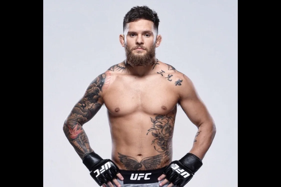
Diagnosed with moyamoya in 2023 and underwent surgery at Stanford, following a path strikingly similar to Vince's. His diagnosis brought further attention to the disease within combat sport.
The son of Will Yun Lee (The Good Doctor) was misdiagnosed for a year before receiving the correct moyamoya diagnosis. His story illustrates the cost of low clinical awareness.
The actress and singer, best known for "This Is Me," suffered a stroke in 2018 connected to moyamoya disease. Her experience underscores that the condition can affect anyone at any stage of life.
The information on this page is for awareness and education and is not a substitute for professional medical advice, diagnosis, or treatment. If you suspect you or someone you know may have moyamoya disease, consult a neurologist or cerebrovascular specialist. See our editorial policy for how we source and review content.
Moyamoya is a rare progressive cerebrovascular condition in which the arteries at the base of the brain narrow and close. The body responds by growing a tangled web of fragile collateral vessels that look like a puff of smoke on imaging. About 1 in 100,000 people in the US are affected.
Moyamoya (もやもや) is Japanese for puff of smoke or hazy. Japanese neurosurgeons Suzuki and Takaku coined the term in 1969 to describe the wispy collateral vessels visible on cerebral angiography. The Japanese name is now used worldwide.
Moyamoya affects roughly 1 in 100,000 people. It is approximately ten times more common in East Asian populations than in the US, and women are affected about twice as often as men. The disease peaks in two age groups, 5 to 10 years old and 30 to 50 years old.
No. Moyamoya causes strokes but is itself a chronic narrowing of the brain's arteries. Without treatment, around 40% of patients have another stroke within 5 years. Bypass surgery dramatically reduces that risk by restoring blood flow around the blockage.
There is no cure. Surgical bypass restores blood flow and reduces stroke risk, but the underlying narrowing remains. Patients need lifelong follow-up imaging to monitor vessel health and the success of the bypass. May 6 is recognised as Moyamoya Awareness Day.
Awareness saves lives. Research brings us closer to a cure. Share this page with someone who needs it, support the cause, or get involved with Miles for Moyamoya.